The effect of SSRIs on threat of shock potentiated neural circuitry
Study code
NBR45
Lead researcher
Dr Oliver Robinson
Study type
Participant re-contact
Institution or company
University College London
Researcher type
Academic
Speciality area
Mental Health
Summary
This is an experimental research study designed to examine the effects of antidepressants on emotional processing and underlying neural circuitry.
This study will use a between-subject double-blind design: participants will be randomly allocated to take 14-21 days of daily escitalopram(10mg) or placebo. The first session will be a screening session lasting around 1.5 hours where participants will be asked to complete standardised structured interviews and complete some self-report questionnaires to determine their cognitive and emotional traits.
We are recruiting two groups of participants: individuals with high anxiety symptoms, and control individuals with no personal or family history of mood and anxiety disorders.
We are requesting access to the GLAD study solely to recruit the former group. If participants meet the inclusion/exclusion criteria of this study and are willing to take part, they will complete two sessions of computerised cognitive tasks in a magnetic resonance imaging scanner(before and after the course of medication).This allows the impact of the intervention on the neural circuitry involved in processing emotional stimuli to be examined.
These sessions will last approximately 2.5hours. Some tasks will have periods of ‘threat of shock’ which serve to induce temporary stress, and matched safe periods.
Shock levels will be individually calibrated to find appropriate levels of shock for each participant. The shocks are rare and harmless, and comparable to a rubber band being snapped against the wrist/ankle–this procedure has been used in multiple previous projects with no consequent adverse events.
Between the two testing sessions, participants will be given either escitalopram or placebo tablets to take home. They will be given an information pack and clinical cover for the duration of the study.
Research publication
Lukow, P. B., Lowther, M., Pike, A. C., Yamamori, Y., Chavanne, A. V., Gormley, S., Aylward, J., McCloud, T., Goble, T., Rodriguez-Sanchez, J., Tuominen, E. W., Buehler, S. K., Kirk, P., & Robinson, O. J. (2024). Amygdala activity after subchronic escitalopram administration in healthy volunteers: A pharmaco-functional magnetic resonance imaging study. Journal of psychopharmacology (Oxford, England), 38(12), 1071–1082. https://doi.org/10.1177/02698811241286773
Research findings
Background: Selective serotonin reuptake inhibitors (SSRIs) are used for the treatment of several conditions including anxiety disorders, but the basic neurobiology of serotonin function remains unclear. The amygdala and prefrontal cortex are strongly innervated by serotonergic projections and have been suggested to play an important role in anxiety expression. However, serotonergic function in behaviour and SSRI-mediated neurobiological changes remain incompletely understood.
Aims: To investigate the neural correlates of subchronic antidepressant administration.
Methods: We investigated whether the 2- to 3-week administration of a highly selective SSRI (escitalopram) would alter brain activation on a task robustly shown to recruit the bilateral amygdala and frontal cortices in a large healthy volunteer sample. Participants performed the task during a functional magnetic resonance imaging acquisition before (n = 96) and after subchronic escitalopram (n = 46, days of administration mean (SD) = 15.7 (2.70)) or placebo (n = 40 days of administration mean (SD) = 16.2 (2.90)) self-administration.
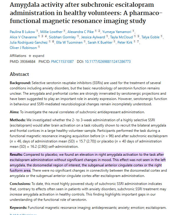
Results: Compared to placebo, we found an elevation in right amygdala activation to the task after escitalopram administration without significant changes in mood. This effect was not seen in the left amygdala, the dorsomedial region of interest, the subgenual anterior cingulate cortex or the right fusiform area. There were no significant changes in connectivity between the dorsomedial cortex and amygdala or the subgenual anterior cingulate cortex after escitalopram administration.
Conclusions: To date, this most highly powered study of subchronic SSRI administration indicates that, contrary to effects often seen in patients with anxiety disorders, subchronic SSRI treatment may increase amygdala activation in healthy controls. This finding highlights important gaps in our understanding of the functional role of serotonin.
